EU inference
All models run through Cloudflare AI Gateway in front of EU providers. Zero data retention where supported.
For thirty years, microscopy analysis has lived in a graveyard of incompatible plugins, dead Python environments, and macros that break the moment lighting changes.
Orasis — from the Ancient Greek ὄρᾰσις, "the act of seeing" — brings the best open-source engines under one roof: Cellpose, Fiji, scikit-image, ilastik, MyoFuse. Then puts an AI on top that actually understands what you're looking at.
Drag-and-drop ingestion for imaging files up to 4 GB. Natively reads .czi, .tif, .ome.tiff, .lif, and .nd2 — no conversion, no plugins.
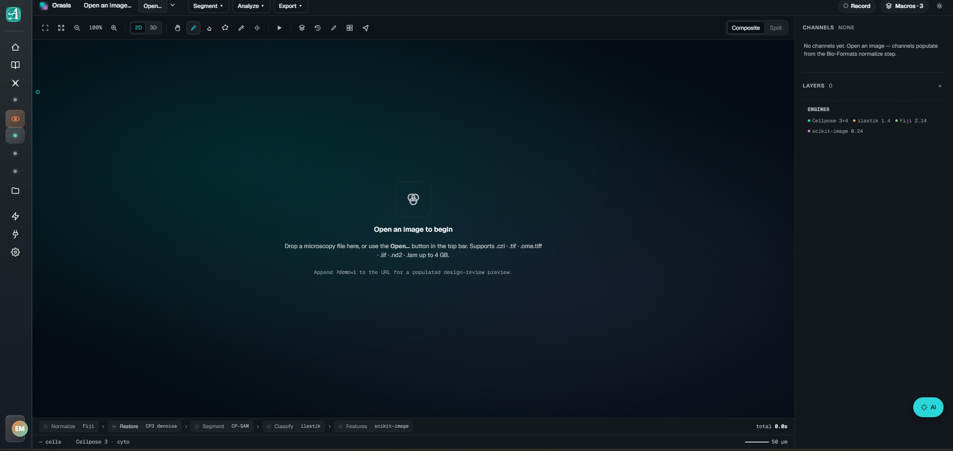
Orasis unifies industry-leading algorithms under a single, cohesive interface, streamlining complex analytical workflows.
Core processing and rigorous quantitative feature extraction (v2.14 & v0.24).
Advanced pixel classification via interactive Machine Learning (v1.4).
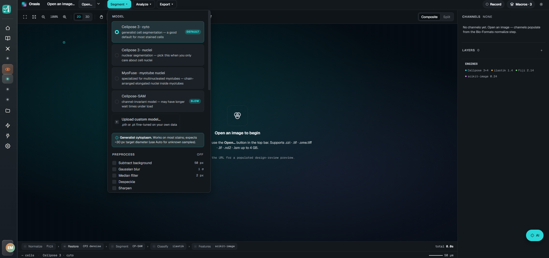
State-of-the-art deep learning models for generalist cyto and nuclei segmentation.
A robust foundation model capable of segmenting diverse biological images with unprecedented generalisation and minimal fine-tuning.
A highly specialised plugin engineered specifically for the analysis of multinucleated myotubes and internal nuclei positioning.
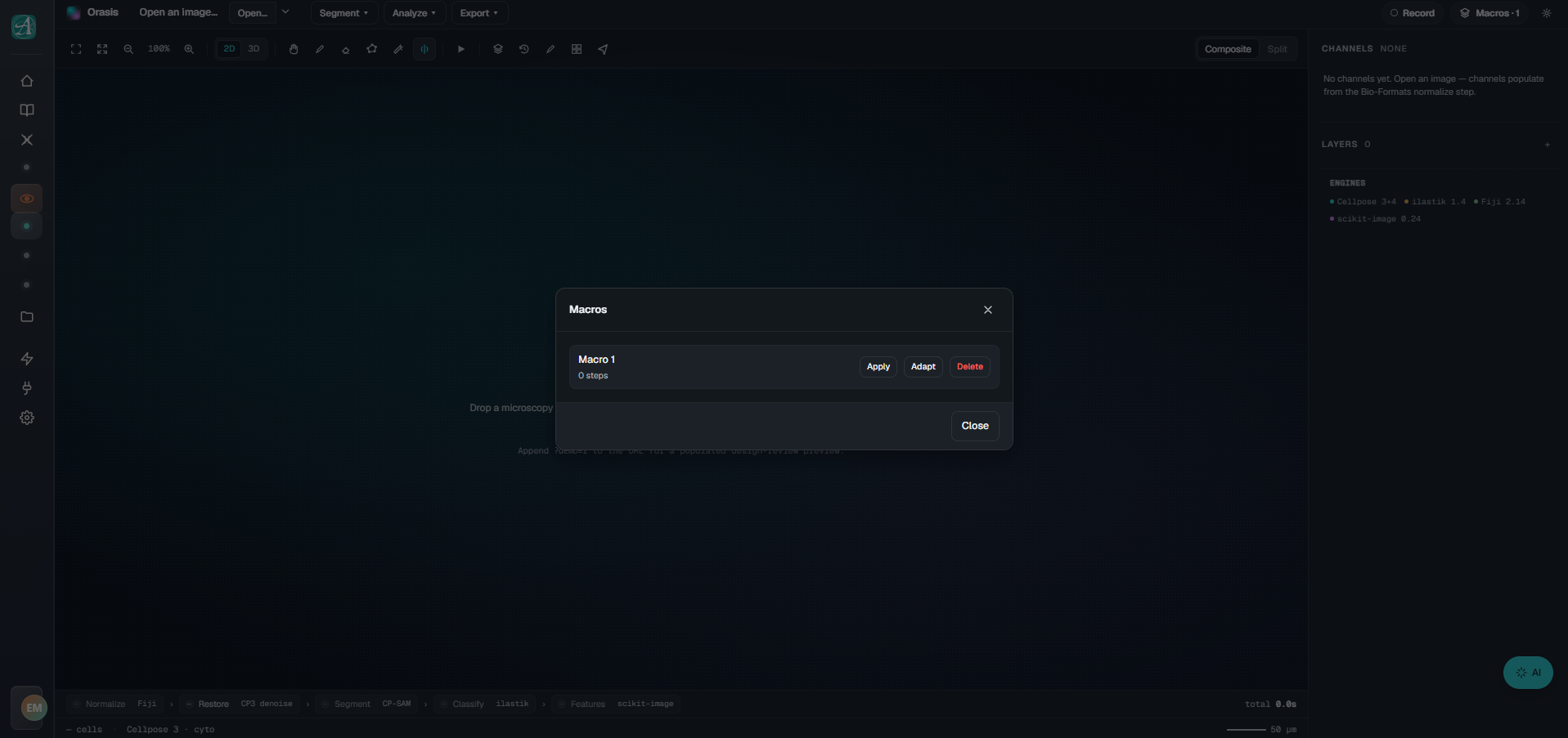
Traditional image analysis relies on static macros — rigid scripts that break the moment lighting, magnification, or sample density shifts. Orasis still lets you save and replay macros, but with one decisive difference: the AI adapts them.
Hit "Adapt" and Orasis looks at the new image — contrast gradients, morphological variance, signal-to-noise — and re-tunes the pipeline on the fly. Same intent, new sample, accurate result. No more rerunning a macro on twenty fields just to discover the threshold drifted on field three.
And when you don't want to click at all, there's the chat. Type what you want. The AI sees the image, picks the right engine, adjusts parameters, executes, and shows its work. "Segment the nuclei, count them, flag anything that looks apoptotic." That's the command.
A vision-language model that actually helps — one that lives next to your data, reads the image, and has hands on the segmentation engines. Suggestions are sourced, parameters are visible, the final call is always yours.
Not every analysis happens at a workstation. Half of microscopy lives at the bench — through an eyepiece, under a gel doc, holding a phone in one hand and a pipette in the other. So we built for that too.
Point your phone through the eyepiece. Snap. The image is sent to our EU inference cluster, where Cellpose SAM does the heavy lifting, and the segmented result lands back on your screen in seconds — confluence, cell counts, the lot.
For cell-culture checks, passaging decisions, and the "is this confluent enough yet?" question you ask twelve times a day. No laptop required.
Photograph a Western blot or DNA gel on your phone — angle, glare, fingerprints and all. The image is processed on our EU inference cluster: perspective corrected, lanes and bands identified, quantification returned with confidence intervals. Results land on your phone in seconds.
Built on the same lineage as GelGenie and the open-source gel-analysis community, with a careful AI layer on top. The reading is sourced. The disagreement is yours.
All models run through Cloudflare AI Gateway in front of EU providers. Zero data retention where supported.
We don't fine-tune on your data. The same model the next user gets is the one you're using today.
Confidence is shown. Disagreement is logged. The note is yours, not the model's.
One tap to flag a Biovision answer as wrong. We log it; the next reading benefits from your correction.
Orasis doesn't replace the tools you already trust — it stitches them together and adds an AI that can drive them. Every engine below is the work of researchers and maintainers who have given their code to the community. Thank you.
Biovision rolls out as part of the Aspis Bio alpha. Drop your email to be notified when your spot is ready.